Monday, August 30, 2010

Inherited Heart Disease

Ellis-van Creveld Syndrome

Introduction

Background

  • Richard W.B. Ellis of Edinburgh and Simon van Creveld of Amsterdam first described Ellis–van Creveld (EVC) syndrome. They met in a train compartment while traveling to a pediatrics conference in England in the late 1930s and discovered that each had a patient with the syndrome. In 1940, Ellis and van Creveld (Ellis and van Creveld, 1940) formally described the syndrome that would bear their names, although they termed it chondroectodermal dysplasia.
  • Disproportionate dwarfism, postaxial polydactyly, ectodermal dysplasia, a small chest, and a high frequency of congenital heart defects characterize this autosomal recessive syndrome, which has increased incidence among persons of Old Order Amish descent.
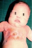
Newborn with Ellis–van Creveld syndrome. No... Newborn with Ellis–van Creveld syndrome. Note the narrow chest.


Postaxial polydactyly


Natal teeth and lip tie.


Pathophysiology

  • Pathophysiology is unknown; however, recent identification of the EVC gene should lead to a better understanding.
  • Histopathologic examination of fetuses with Ellis–van Creveld syndrome revealed that the cartilage of long bones showed chondrocyte disorganization in the physeal growth zone. Variable chondrocyte disorganization was seen in the central physeal growth zone of the vertebrae.

Frequency

United States

  • In the general population, the frequency is 1 case per 60,000 live births.
  • Among persons from the Old Order Amish, the incidence is estimated at 5 cases per 1000 live births.
  • The frequency of carriers in this population may be as high as 13%.

Mortality/Morbidity

  • Thoracic dysplasia leads to respiratory insufficiency and cardiac anomalies lead to death in infancy in 50% of patients.
  • Patients who survive infancy have a normal life expectancy.

Race

  • The highest frequency of Ellis–van Creveld syndrome is seen in one particular inbred population, the Old Order Amish community in Lancaster County, Pennsylvania, where the largest pedigree has been described (52 cases in 30 sibships).
  • Among the Amish, the abnormal gene can be traced to the immigrants Samuel King and his wife, whose identity is not known.
  • No other ethnic group has a high incidence of Ellis–van Creveld syndrome.

Sex & Age

  • Frequency of Ellis–van Creveld syndrome is equal in males and females.
  • In patients with Ellis–van Creveld syndrome, physical findings, such as disproportionate extremities, small stature, polydactyly, cardiac defects, and minor dysmorphic features, are seen at birth.

Clinical History

  • In the prenatal period, intrauterine growth retardation, skeletal malformations, and cardiac defects can be depicted on ultrasound images in fetuses with Ellis–van Creveld (EVC) syndrome.
  • Family history may include parental consanguinity or previously affected siblings or family members.
  • Neonatal history may include small size at birth, slow growth, and skeletal anomalies are the initial symptoms. Natal teeth may be present.
  • Heart disease may be manifested as failure to thrive, cyanosis, shortness of breath, cardiac murmur, or other signs suggestive of heart failure.
  • Developmentally, most patients have had intelligence in the normal range. Occasionally, patients present with associated brain malformations and developmental delay.

Physical

  • The variable phenotype affects multiple organs.
  • A clinical tetrad of Ellis–van Creveld syndrome consists of chondrodystrophy, polydactyly, ectodermal dysplasia, and cardiac anomalies.
    • Chondrodystrophy (the most common feature affecting the tubular bones)
      • Disproportionate dwarfism (small stature of prenatal onset; average adult height, 109-155 cm)
      • Progressive distal limb shortening, symmetrically affecting the forearms and lower legs
    • Polydactyly (constant findings)
      • Bilateral and postaxial
      • Polydactyly, observed in the hands in most cases but in the feet in 10% of cases
    • Hidrotic ectodermal dysplasia (observed in as many as 93% of cases)
      • Nails are hypoplastic, dystrophic, and friable. Nails can be completely absent in some cases.
      • Tooth involvement may include neonatal teeth, partial anodontia, small teeth, and delayed eruption. Enamel hypoplasia may result in abnormally shaped teeth with frequent malocclusion.
      • Hair may occasionally be sparse.
    • Congenital cardiac anomalies
      • Heart defects occur in 50-60% of patients; the most common anomaly is a common atrium (40%).
      • Other cardiac anomalies include atrioventricular canal, ventricular septal defect, atrial spetal defect, and patent ductus arteriosus.
      • The cardiac anomaly is the major cause of shortened life expectancy.
  • Other anomalies may also be present.
    • Musculoskeletal anomalies include low-set shoulders, a narrow thorax frequently leading to respiratory difficulties, knock knees, lumbar lordosis, broad hands and feet, and sausage-shaped fingers.
    • Oral lesions include the following:
      • A fusion of the anterior portion of the upper lip to the maxillary gingival margin, resulting in an absence of mucobuccal fold and the upper lip to present a slight V-notch in the middle
      • Short upper lip, bound by frenula to alveolar ridge (lip tie)
      • Often serrated lower alveolar ridge
      • Teeth may be prematurely erupted at birth or exfoliate prematurely
    • Occasional genitourinary anomalies include hypospedias, epispadias, hypoplastic penis, cryptorchidism, vulvar atresia, focal renal tubular dilation in medullary region, nephrocalcinosis, renal agenesis, and megaureters.
    • Occasionally, CNS anomalies or mental retardation are present.
  • Clinical manifestations in heterozygous carriers
    • Polydactyly has been reported in relatives of 4 unrelated Ellis–van Creveld syndrome families.
    • A father of a child with Ellis–van Creveld syndrome who had finger and teeth abnormalities has been reported, as have several other reports of symptomatic heterozygous manifestations.
    • The Weyers acrofacial dysostosis, an autosomal dominant disorder described in 1952, is characterized by variable extremities and facial features. This condition has been found to be associated with EVC and EVC2 mutations that have confirmed that Weyers dysostosis represents the heterozygous expression of the mutation that causes Ellis–van Creveld syndrome.

Causes

  • Ellis–van Creveld syndrome has an autosomal recessive inheritance.
  • The EVC gene has been mapped to chromosome band 4p16 using linkage analysis of 9 interrelated Amish pedigrees and 3 unrelated families from Mexico, Ecuador, and Brazil. A 992 amino acid protein encoded by this gene is predicted to contain a leucine zipper domain, 3 putative nuclear localization signals, and a putative transmembrane domain.
  • Mutations in the EVC gene were identified in patients with Ellis–van Creveld syndrome.
  • Ellis–van Creveld syndrome is also caused by mutations in a second gene, called EVC2, that gives rise to the same phenotype of the syndrome.
  • Patients with Weyers acrodental dysostosis were also found to have mutations in the gene, which confirms that Ellis–van Creveld syndrome and Weyers dysostosis are allelic.

Prognosis

  • Approximately 50% of patients with Ellis–van Creveld (EVC) syndrome die in early infancy as a consequence of cardiorespiratory problems. Most survivors have intelligence in the normal range.
  • Final adult height is 43-60 inches.
  • Usually, some limitation of hand function is observed, such as inability to form a clenched fist.
  • Dental problems are frequent.
  • End-organ involvement may include the following:
    • Renal involvement including nephrotic syndrome, nephronophthisis, and renal failure
    • Hepatic involvement, including a congenital paucity of bile ducts that leads to progressive fibrosis and hepatic failure
    • Hematologic involvement ranges from myelodysplastic changes with dyserythropoiesis to acute leukemia.

Treatment

Medical Care

  • The management of Ellis–van Creveld (EVC) syndrome is multidisciplinary.
  • Care for respiratory distress, recurrent respiratory infections, and cardiac failure is supportive.
  • Dental care in childhood includes the following:
    • Neonatal teeth should be removed because they may impair feeding.
    • Prevention of caries includes dietary counseling, plaque control, and oral hygiene instruction.
    • Crown or composite build-ups for microdonts may be indicated.
    • Partial dentures can maintain space and improve mastication, esthetics, and speech due to congenitally missing teeth.
    • Orthodontic treatment is needed for bone deformity, especially knee valgus with depression of the lateral tibial plateau and dislocation of the patella.
  • For dental care during adulthood, implants and prosthetic rehabilitation are required to replace congenitally missing teeth.
  • Short stature is considered resulting of chondrodysplasia of the legs and the possible treatment with growth hormone is considered ineffective, unless the patient is also deficient in growth hormone.

Surgical Care

  • Orthopedic procedures correct polydactyly and other orthopedic malformations.
  • Cardiac surgery may be needed to correct cardiac anomalies.
  • Thoracic expansion has been attempted in some patients.
  • Dental care is usually necessary.
  • Urologic surgery is required if epispadias, cryptorchidism, or both are present.
  • Perioperative morbidity may result from difficulties with airway management and pulmonary abnormalities.15
    • Although these concerns are less common than congenital heart disease, abnormalities leading to difficulties in airway management include cleft lip and plate orodental malformations.
    • Frenulae, or fusions between the inner upper lip and gum, as well as maxillary or mandibular deformities, may lead to difficulties in bag-valve-mask ventilation and should be identified during the preoperative evaluation.
    • Dental abnormalities, such as peg teeth or natal teeth, may be more prone to dislodgement during airway instrumentation.
    • A single report describes a patient with Ellis–van Creveld syndrome who presented with congenital stridor related to a cyst involving the neck and airway.

Consultations

  • Clinical geneticist
  • Cardiologist
  • Pulmonologist
  • Orthopedist
  • Urologist
  • Physical and occupational therapist
  • Dentist
  • Psychologist
  • Developmental pediatrician (if developmental delay is present)
  • Pediatric neurologist (if developmental delay is present)

Diet

  • No special diet is required unless cardiac failure necessitates dietary restrictions.

Activity

  • Activities may be limited secondary to cardiorespiratory status or skeletal anomalies.

Medication

Specific drug therapy is not currently a component of the standard of care for Ellis–van Creveld (EVC) syndrome. Treat systemic sequelae as needed.

Differential Diagnoses

Other Problems to Be Considered

  • Other short rib polydactyly syndromes include the following:
    • Saldino-Noonan syndrome (type I)
    • Majewski syndrome (type II)
    • Verma-Naumoff syndrome (type III)
    • Beemer-Langer syndrome (type IV)
    • Asphyxiating thoracic dystrophy (Jeune syndrome)
  • Polydactyly and hypodontia have been described in Weyers acrodental dysostosis, which is allelic with EVC and in trisomy 13. Weyers acrodental dysostosis is an autosomal dominant condition that is the heterozygous manifestation of the EVC gene; disproportionate dwarfism, heart defect, and thoracic dysplasia are not present.

Workup

Laboratory Studies

  • Sequencing of EVC and EVC2 identified mutations in two thirds of patients with Ellis-van Creveld (EVC) syndrome.
  • Gene testing for mutational analysis of EVC and EVC2 is not currently available clinically.

Imaging Studies

  • A skeletal survey is necessary to define skeletal anomalies. Expected findings include the following:
    • Acromesomelia (relative shortening of the distal and middle segment of the limbs) - Most prominent in the hands, where the distal and middle phalanges are shorter than the proximal phalanx
    • Polydactyly (ulnar side)
    • Multiple varieties of carpal fusion
    • Small iliac crests and sciatic notches (may be revealed on pelvic radiographs)
    • Valgus deformity of the knee
    • Fibula disproportionately smaller than the tibia
    • Thorax (short ribs, narrow)
    • Retarded bone maturation
    • Other findings - Fusion of the hamate and capitate bones of the wrist, cubitus valgus, hypoplastic cubitus, supernumerary carpal bone center, clinodactyly of the 5th finger
  • Chest radiography, ECG, and echocardiography (to evaluate cardiac anatomy) are indicated.
  • Head MRI may infrequently reveal brain anomalies.
  • Renal ultrasonography may infrequently reveal renal anomalies.

Other Tests

  • Consider eye examination to exclude eye anomalies, which have been infrequently described.

Histologic Findings

  • Disorganization of chondrocytes in the physeal growth zone of the long bones and vertebrae in the prenatal period and retardation of physeal growth zones in childhood.

Congenital Heart Diseases


Congenital Heart Diseases

A baby's heart begins to develop at conception, but is completely formed by eight weeks into the pregnancy. Congenital heart defects happen during this crucial first eight weeks of the baby's development. Specific steps must take place in order for the heart to form correctly. Often, congenital heart defects are a result of one of these crucial steps not happening at the right time, leaving a hole where a dividing wall should have formed, or a single blood vessel where two ought to be, for example.

Causes of congenital heart disease

The vast majority of congenital heart defects have no known cause. Mothers will often wonder if something they did during the pregnancy caused the heart problem. In most cases, nothing can be attributed to the heart defect. Some heart problems do occur more often in families, so there may be a genetic link to some heart defects. Some heart problems are likely to occur if the mother had a disease while pregnant and was taking medications, such as anti-seizure medicines. However, most of the time, there is no identifiable reason as to why the heart defect occurred.

Congenital heart problems range from simple to complex. Some heart problems can be watched by the baby's physician and managed with medicines, while others will require surgery, sometimes as soon as in the first few hours after birth. A baby may even "grow out" of some of the simpler heart problems, such as patent ductus arteriosus (PDA) or atrial septal defect (ASD), since these defects may simply close up on their own with growth. Other babies will have a combination of defects and require several operations throughout their lives.

Types of Congenital heart diseases:

  • Artrial Septal Defect
  • Ventrical Septal Defect
Artrial Septal Defect

An atrial septal defect (ASD) — sometimes referred to as a hole in the heart — is a type of congenital heart in which there is an abnormal opening in the dividing wall between the upper filling chambers of the heart (the atria). In most cases ASDs are diagnosed and treated successfully with few or no complications.

Kids with an atrial septal defect (ASD) have an opening in the wall (septum) between the atria. As a result, some oxygenated blood from the left atrium flows through the hole in the septum into the right atrium, where it mixes with oxygen-poor blood and increases the total amount of blood that flows toward the lungs. The increased blood flow to the lungs creates creates a swishing sound, known as a heart murmur. This heart murmur, along with other specific heart sounds that can be detected by a cardiologist, may be clues that a child has an ASD.

ASDs can be located in different places on the atrial septum, and they can be different sizes. The symptoms and medical treatment of the defect will depend on those factors. In some rare cases, ASDs are part of more complex types of congenital heart disease. It's not clear why, but ASDs are more common in girls than in boys.

Figure A shows the normal structure and blood flow in the interior of the heart. Figure B shows a heart with an atrial septal defect. The hole allows oxygen-rich blood from the left atrium to mix with oxygen-poor blood from the right atrium.

Causes

ASDs occur during fetal development of the heart and are present at birth. During the first weeks after conception, the heart develops. If a problem occurs during this process, a hole in the atrial septum may result.

In some cases, the tendency to develop a ASD may be genetic. There can be genetic syndromes that cause extra or missing pieces of chromosomes that can be associated with ASD. For the vast majority of children with a defect, however, there's no clear cause of the ASD.

Signs and Symptoms

The size of an ASD and its location in the heart will determine what kinds of symptoms a child experiences. Most children who have ASDs seem healthy and appear to have no symptoms. Generally, kids with an ASD feel well and grow and gain weight normally. Infants and children with larger, more severe ASDs, however, may possibly show some of the following signs or symptoms:

  • poor appetite
  • poor growth
  • fatigue
  • shortness of breath
  • lung problems and infections, such as pneumonia

If an ASD is not treated, health problems can develop later, including an abnormal heart rhythm (known as an atrial arrhythmia) and problems in how well the heart pumps blood. As kids with ASDs get older, they may also be at an increased risk for stroke, since a blood clot that develops can pass through the hole in the wall between the atria and travel to the brain. Pulmonary hypertension (high blood pressure in the lungs) may also develop over time in older patients with larger untreated ASDs.

Fortunately, most kids with ASD are diagnosed and treated long before the heart defect causes physical symptoms. Because of the complications that ASDs can cause later in life, pediatric cardiologists often recommend closing ASDs early in childhood.

Diagnosis

Generally, a child's doctor hears the heart murmur caused by ASD during a routine checkup or physical examination. ASDs are not always diagnosed as early in life as other types of heart problems, such as ventricular septal defect (a hole in the wall between the two ventricles). The murmur caused by an ASD is not as loud and may be more difficult to hear than other types of heart murmurs, so it may be diagnosed any time between infancy and adolescence (or even as late as adulthood).

If a doctor hears a murmur and suspects a heart defect, the child may be referred to a pediatric cardiologist, a doctor who specializes in diagnosing and treating childhood heart conditions. If an ASD is suspected, the cardiologist may order one or more of the following tests:

  • chest X-ray, which produces an image of the heart and surrounding organs
  • electrocardiogram (EKG), which records the electrical activity of the heart and can indicate volume overload of the right side of the heart
  • echocardiogram (echo), which uses sound waves to produce a picture of the heart and to visualize blood flow through the heart chambers. This is often the primary tool used to diagnose ASD.

Treatment

Once an ASD is diagnosed, treatment will depend on the child's age and the size, location, and severity of the defect. In kids with very small ASDs, the defect may close on its own. Larger ASDs usually won't close, and must be treated medically. Most of these can be closed in a cardiac catheterization lab, although some ASDs will require open-heart surgery.

A child with a small defect that causes no symptoms may simply need to visit a pediatric cardiologist regularly to ensure that there are no problems; often, small defects will close spontaneously without any treatment during the first years of life. In general, a child with a small ASD won't require restrictions on his or her physical activity.

In most children with ASD, though, doctors must close the defect if it has not closed on its own by the time a child is old enough to start school.

Depending on the position of the defect, many children with ASD can have it corrected with a cardiac catheterization. In this procedure, a thin, flexible tube called a catheter is inserted into a blood vessel in the leg that leads to the heart. A cardiologist guides the tube into the heart to make measurements of blood flow, pressure, and oxygen levels in the heart chambers. A special implant can be positioned into the hole in the septum. The device is designed to flatten against the septum on both sides to close and permanently seal the ASD. In the beginning, the natural pressure in the heart holds the device in place. Over time, the normal tissue of the heart grows over the device and covers it entirely. This non-surgical technique for closing an ASD eliminates the scar on the chest needed for the surgical approach, and has a shorter recovery time, usually just an overnight stay in the hospital.

Because there is a small risk of blood clots forming on the closure device while new tissue heals over it, children who undergo device closure of an ASD may need to be on medications for several months after the procedure to prevent clots from forming.

If surgical repair for ASD is necessary, a child will undergo open-heart surgery. In this procedure, a surgeon makes a cut in the chest and a heart-lung machine is used to do the work of the circulation while the heart surgeon closes the hole. The ASD may be closed directly with stitches or by sewing a patch of surgical material over the defect. Eventually, the tissue of the heart heals over the patch or stitches, and by 6 months after the surgery, the hole will be completely covered with tissue.

For 6 months following catheterization or surgical closure of an ASD, antibiotics are recommended before routine dental work or surgical procedures to prevent infective endocarditis. Once the tissue of the heart has healed over the closed ASD most people who have had their ASDs corrected no longer need to worry about having a higher risk of infective endocarditis.

Your doctor will discuss other possible risks and complications with you prior to the procedure. Typically, after repair and adequate time for healing, children with ASD rarely experience further symptoms or disease.

Ventrical Septal Defect

A ventricular septal defect (VSD) — sometimes referred to as a hole in the heart — is a type of congenital heart defect in which there is an abnormal opening in the dividing wall between the main pumping chambers of the heart (the ventricles). VSDs are the most common congenital heart defect, and in most cases they're diagnosed and treated successfully with few or no complications.

The right and left-sided pumping chambers (ventricles) are separated by shared wall, called the ventricular septum.

Kids with a VSD have an opening in this wall. As a result, when the heart beats, some of the blood in the left ventricle (which has been enriched by oxygen from the lungs) is able to flow through the hole in the septum into the right ventricle. In the right ventricle, this oxygen-rich blood mixes with the oxygen-poor blood and goes back to the lungs. The blood flowing through the hole creates an extra noise, which is known as a heart murmur. The heart murmur, can be heard when a doctor listens to the heart beat with a stethoscope.

Figure A shows the normal anatomy and blood flow of the interior of the heart. Figure B shows two common locations of ventricular septal defects. The defect allows oxygen-rich blood from the left ventricle to mix with oxygen-poor blood in the right ventricle.


VSDs can be located in different places on the ventricular septum, and they can be different sizes. The symptoms and medical treatment of the VSD will depend on those factors. In some rare cases, VSDs are part of more complex types of congenital heart disease.

What Causes a VSD?

Ventricular septal defects occur during fetal heart development and are present at birth. During the first weeks after conception, the heart develops from a large tube, dividing into sections that will eventually become the walls and chambers. If a problem occurs during this process, it can create a hole in the ventricular septum.

In some cases, the tendency to develop a VSD may be genetic. There can be genetic syndromes that cause extra or missing pieces of chromosomes that can be associated with VSD. For the vast majority of children with a defect, however, there's no clear cause of the VSD.

Signs and Symptoms of a VSD

VSDs are usually found in the first few weeks of life by a doctor during a routine checkup. The doctor will be able to detect a heart murmur, which is due to the sound of blood as it passes between the left and right ventricles. The murmur associated with a VSD has certain features that allow a doctor to distinguish it from heart murmurs due to other causes.

The size of the hole and its location within the heart will determine whether VSD causes any symptoms. Small VSDs will not typically cause any symptoms, and may ultimately close on their own. Older kids or teens who have small VSDs that persist usually don't experience any symptoms other than the heart murmur that doctors hear. They may need to see a doctor regularly to check on the heart defect and make sure it isn't causing any problems.

Moderate and large VSDs that haven't been treated in childhood may cause noticeable symptoms. Babies may have faster breathing and get tired out during attempts to feed. They may start sweating or crying with feeding, and may gain weight at a slower rate.

These signs generally indicate that the VSD will not close by itself, and cardiac surgery may be needed to close the hole. Surgery is typically done within the first 3 months of life to prevent it from causing other complications. A cardiologist can prescribe medication to lessen symptoms before surgery.

People with VSD are at greater risk in their lifetime of developing endocarditis, an infection of the inner surface of the heart. This occurs when bacteria in the bloodstream infect the lining of the heart. Bacteria are always in our mouths, and small amounts are introduced into the bloodstream when we chew and brush our teeth. The best way to protect the heart from endocarditis is to to reduce oral bacteria by brushing and flossing daily, and visiting the dentist regularly. In general, it is not recommended that patients with simple VSDs take antibiotics before dental visits, except for the first 6 months after surgery.

Diagnosing a VSD

If your child is discovered to have a heart murmur that was not noticed earlier, a doctor may refer you to a pediatric cardiologist, a doctor who specializes in diagnosing and treating childhood heart conditions.

In addition to doing a physical exam, the cardiologist take your child's medical history. If a VSD is suspected, the cardiologist may order one or more of the following tests:

  • a chest X-ray, which produces a picture of the heart and surrounding organs
  • an electrocardiogram, which records the electrical activity of the heart
  • an echocardiogram (echo), which uses sound waves to produce a picture of the heart and to visualize blood flow through the heart chambers. This is often the primary tool used to diagnose VSD.
  • a cardiac catheterization, which provides information about the heart structures as well as blood pressure and blood oxygen levels within the heart chambers. This test is usually performed for VSD only when additional information is needed that other tests cannot provide.

Treating a VSD

Once an VSD is diagnosed, treatment will depend on the child's age and the size, location, and severity of the defect. A child with a small defect that causes no symptoms may simply need to visit a cardiologist regularly to make sure that there are no other problems. In most children, a small defect will close on its own without surgery. Some may not close, but they do not get any larger. In cases where the VSD is small and has not closed, there are generally no restrictions to activities or to playing sports.

For kids with medium to large VSDs, surgery may be necessary. In most cases, this takes place within the first few weeks to months of life. In this procedure, the surgeon makes an incision in the chest wall, and a heart-lung machine is used to do the work of the circulation while the surgeon closes the hole. The surgeon can stitch the hole closed directly or, more commonly, sew a patch of manmade surgical material over it. Eventually, the tissue of the heart heals over the patch or stitches, and by 6 months after the surgery, the hole will be completely covered with tissue.

Certain types of VSDs may be closed during cardiac catheterization. A thin, flexible tube called a catheter is inserted into a blood vessel in the leg that leads to the heart. A cardiologist guides the tube into the heart to make measurements of blood flow, pressure, and oxygen levels in the heart chambers. A special implant, shaped into two disks formed of flexible wire mesh, can be positioned into the hole in the septum. The device is designed to flatten against the septum on both sides to close and permanently seal the VSD.

After healing from surgery or catheterization, kids with VSDs are considered cured and should have no further symptoms or problems.

Sunday, August 29, 2010

General Heart Diseases


General Heart Diseases

  • Myocardial Infarction (Heart Attack)
    • Atherosclerosis
    • Coronary Thrombosis
  • Aortic Dissection
  • Cardiac Arrhythmia (Irregular Heart Beat, or Arrhythmia)
  1. Myocardial Infarction (Heart Attack)

The proper use of the non-medical term "heart attack" is "Myocardial Infarction". Either term is scary. "Myocardial Infarction" (abbreviated as "MI") means there is death of some of the muscle cells of the heart as a result of a lack of supply of oxygen and other nutrients. This lack of supply is caused by closure of the artery ("coronary artery") that supplies that particular part of the heart muscle with blood. This occurs 98% of the time from the process of arteriosclerosis ("hardening of the arteries") in coronary vessels.


Although it once was felt that most heart attacks were caused from the slow closure of an artery, say from 90 or 95% to 100%, it is now clear that this process can occur in even minor blockages where there is rupture of the cholesterol plaque. This in turn causes blood clotting within the artery, blocking the flow of blood. This sort of event is illustrated above. The heart muscle which is injured in this way can cause irregular rhythms which can be fatal, even when there is enough muscle left to pump plenty of blood. When the injured area heals, it will leave a scar. While the heart won't be able to pump quite as much as before, there is often plenty of good muscle left to take care of the job, and recovery can be quite complete.

While heart attacks are clearly scary, with modern techniques, patients survive most of them. Furthermore, most can have a long and satisfying life, perhaps more satisfying than before.


Causes

Predisposing factors include:

❑ positive family history

❑ hypertension

❑ smoking

❑ elevated levels of serum triglycerides, total cholesterol, and low-density lipoproteins

❑ diabetes mellitus

❑ obesity or excessive intake of saturated fats, carbohydrates, or salt

❑ sedentary lifestyle

❑ aging

❑ stress or a type A personality (aggressive, ambitious, competitive, addicted to work, chronically impatient)

❑ drug use, especially cocaine.

Men and postmenopausal women are more susceptible to an MI than premenopausal women, although incidence is rising among females, especially those who smoke and take a hormonal contraceptive.

The site of the MI depends on the vessels involved. Occlusion of the circumflex branch of the left coronary artery causes a lateral wall infarction; occlusion of the anterior descending branch of the left coronary artery, an anterior wall infarction.

True posterior or inferior wall infarctions generally result from occlusion of the right coronary artery or one of its branches. Right ventricular infarctions can also result from right coronary artery occlusion, can accompany inferior infarctions, and may cause right-sided heart failure. With a transmural MI, tissue damage extends through all myocardial layers; with a subendocardial MI, only in the innermost and possibly the middle layers.

Symptoms:

Common Heart Attack Warning Signs

  • Uncomfortable pressure, fullness, squeezing or pain in the center of the chest lasting more than a few minutes
  • Pain spreading to the shoulders, neck or arms.
  • Chest discomfort with lightheadedness, fainting, sweating, nausea or dyspnea (shortness of breath).

Other signs of myocardial infraction may include:

  • Sweating
  • Jaw pain
  • Heartburn or indigestion
  • Upper back pain
  • General feeling of illness

A recent survey reported by the American Heart Association reveals that the majority of American women do not understand the true threat of cardiovascular disease. Despite the fact that heart disease is the leading cause of death among women, a nationwide survey revealed that only 8% of women perceive heart disease as the greatest threat to their health. More than six out of 10 women falsely believe that they are more likely to develop cancer than heart disease.

Diagnosing Myocardial Infarction (Heart Attack)


When symptoms are presented, patients should be evaluated quickly with blood tests and an electrocardiogram. After the patient is stabilized, an echocardiogram and nuclear medicine exam may be performed.

  • Blood work: Blood tests will be performed to detect levels of creatine phosphokinase (CPK), aspartate aminotransferase (AST), lactate dehydrogenase (LDH) and other enzymes released during myocardial infarction.
  • Electrocardiogram (ECG or EKG): An electrocardiogram makes a graphic record of the cardiac activity, either on paper or a computer monitor. An ECG can be beneficial in detecting disease and/or damage.
  • Echocardiogram (heart ultrasound): This diagnostic technique is an excellent first step in investigating congenital heart disease or in evaluating abnormalities of the heart wall. Echocardiography is a non-invasive exam in which images are acquired and viewed in real time without the use of radiation. Echocardiography is often useful in studying the beating heart and provides some information on functional abnormalities of the heart wall, valves and blood vessels. Doppler ultrasound can be used to measure blood flow across a heart valve. Abnormal operation of the valves can be detected by studying the opening and closing function versus normal valve function. Echocardiography may also be used to study congenital heart defects such as a septal defect (a hole in the wall that separates the two chambers of the heart).
  • Nuclear medicine: Nuclear medicine (also called radionuclide scanning) allows visualization of the anatomy and function of an organ. The patient will be given a radionuclide which will assist in the acquisition clear images of the heart with a gamma camera. Nuclear medicine imaging may be used to detect coronary artery disease, myocardial infarction, valve disease, heart transplant rejection, check the effectiveness of bypass surgery, or to select patients for angioplasty or coronary bypass graft.
Duration of a Heart Attack:

A heart attack itself may last several minutes when the symptoms are present. However, because of the damage it causes and the way the heart tries to cope with it, in those people who survive a heart attack, the consequences last a lot longer. This may mean there is a risk of more abnormal heartbeats (arrhythmias) for several hours or days following. For some patients there can be further risk several months later because they may go on to develop heart failure or other problems. This is why special care and medicines are needed for a long time, to reduce the chances of this happening.
  • Following recovery from a heart attack there is damage to the heart muscle, which takes some time to repair.

  • The repair to the heart muscle is not always complete and scarring is usually present.

  • There is always a chance of a recurrence due to the continued presence of diseased coronary arteries that caused the heart attack.

  • There is also the risk of heart failure developing over a period of weeks as the heart reacts to the injury it has sustained.

  • For these reasons it is necessary for patients to be monitored carefully and to receive the appropriate treatment to reduce the risk of further disease progression and other heart attacks.
Treatment of a Heart Attack:

Medical treatment is aimed to open the blocked artery and restore blood flow to the affected area of heart muscle (doctors call this reperfusion). Treatment is also aimed at preventing further damage and the chance of repeat heart attacks in the future.
  • Once the artery is open, the heart attack is generally halted and the patient becomes pain free.

  • The patient is most likely to make a good recovery if reperfusion can be established in the first 4-6 hours of a heart attack.

  • Anti-platelet medicines, for example aspirin, reduce the tendency of platelets (a type of blood cell) in the blood to clump and clot. These medicines help to prevent the arteries from becoming blocked again.

  • Nitroglycerin, a vasodilator (blood vessel dilator), widens the blood vessel by relaxing the muscular wall of the blood vessel.

  • ACE (angiotensin converting enzyme) inhibitors, another type of vasodilator, improve the heart muscle healing process. They do this by blocking the production of a hormone (chemical signal carried in the blood) called angiotensin II.

  • Beta-blocking agents interfere with the nerves controlling the heart by blocking the action of a chemical they release called noradrenaline. They also block a hormone (chemical carried in the blood) called adrenaline. This makes the heart beat more slowly and less forcibly, which decreases the amount of muscle damage and can help to prevent serious arrhythmias.

After a heart attack, many other recommendations may be made including changes in diet, lifestyle, stopping smoking and so on. The aim of these is to try to reduce the chance of having another heart attack. If specific conditions are discovered that have contributed to the heart attack, like high cholesterol or high blood pressure for example, then specific treatments might be needed for these.

Any medical information on this website is not intended as a substitute for informed medical advice and you should not take any action before consulting with a health care professional.

Special considerations


❑ Care for patients who have suffered an MI is directed toward detecting complications, preventing further myocardial damage, and promoting comfort, rest, and emotional well-being. Most MI patients receive treatment in the coronary care unit (CCU), where they’re under constant observation for complications.

❑ On admission to the CCU, record the patient’s blood pressure, temperature, and heart and breath sounds, and monitor them regularly. Also, obtain an ECG.

❑ Assess and record the severity and duration of pain; administer an analgesic. Avoid I.M. injections; absorption from the muscle is unpredictable.

❑ Check the patient’s blood pressure after giving nitroglycerin, especially the first dose.

❑ Frequently monitor the ECG to detect rate changes or arrhythmias. Place rhythm strips in the patient’s chart periodically for evaluation.

❑ During episodes of chest pain, obtain ECG, blood pressure, and pulmonary artery catheter measurements for changes.

❑ Watch for signs and symptoms of fluid retention (crackles, cough, tachypnea, and edema), which may indicate impending heart failure. Carefully monitor daily weight, intake and output, respirations, serum enzyme levels, and blood pressure.

❑ Auscultate for adventitious breath sounds periodically (patients on bed rest frequently have atelectatic crackles, which may disappear after coughing) and for third or fourth heart sounds.

❑ Organize patient care and activities to maximize periods of uninterrupted rest.

❑ Ask the dietary department to provide a clear liquid diet until nausea subsides. A low-cholesterol, low-sodium, caffeine-free diet may be ordered.

❑ Provide a stool softener to prevent straining during defecation, which causes vagal stimulation and may slow the heart rate. Allow use of a bedside commode, and provide as much privacy as possible.

❑ Assist with range-of-motion exercises. If the patient is completely immobilized by a severe MI, turn him often. Antiembolism stockings help prevent venostasis and thrombophlebitis.

❑ Provide emotional support, and help reduce stress and anxiety; administer a tranquilizer, if needed.

❑ Explain procedures, and answer questions. Explaining the CCU environment and routine can ease anxiety. Involve the patient’s family in his care as much as possible.

To prepare the patient for discharge:

❑ Promote adherence measures by thoroughly explaining the prescribed medication regimen and other treatment measures.

❑ Warn the patient about adverse reactions to drugs, and advise him to watch for and report signs and symptoms of toxic reaction (anorexia, nausea, vomiting, and yellow vision, for example, if the patient is receiving digoxin).

❑ Review dietary restrictions with the patient. If he must follow a low-sodium or low-fat and low-cholesterol diet, provide a list of foods that he should avoid. Ask the dietitian to speak to the patient and his family.

❑ Counsel the patient to resume sexual activity progressively.


Wednesday, August 25, 2010

Diseases

In biology, disease refers to any abnormal condition of an organism that impairs function.

The term disease is often used metaphorically for disordered, dysfunctional, or distressing conditions of other things, as in disease of society.